Circulatory System NEB

Circulation:
It is the circulation of internal body fluid through definite vessel through either blood vessels or lymphvessels.
FunctionsoftheCirculatorySystem
1. Respiration-deliversoxygentothecellsandremovingcarbondioxidefromthem.
2. Nutrition-carriesdigestedfoodsubstances tothecellsofthebody.
3. WasteRemoval-disposesofwasteproductsandpoisonsthatwouldharmthebodyiftheyaccumulated.
4. Immunity-helpsprotectthebodyfrom disease.
5. CellularCommunication-thecirculatorysystemprovidesamodeoftransportforhormones.
6.
Thermoregulation-thecirculatorysystemtransportsheat(canbothwarmandcoolbody).
TypesofCirculatory system:
Open Type:
- Blood is not enclosed in blood vessels.
-
It ispumpedintoacavitycalledhemocoelwhereitmixeswithinterstitial fluid.
- - Theinternalorgansaredirectlybathedinblood.
-
Itoccursinorganisimslikesnails,cockroachesandspiders.
ClosedType:
- - In the closed type of circulatory system, the blood
remains inside the blood vessels and does notcome out.
- - Thebloodflowsfromarteriestoveinsthrough smallbloodvessels called capillaries.
- -Itoccurs in organisims likehumans,squids,cats,earthwormsetc.

Typesofcirculation:
1. Singlecirculation:
- Blood flows only once through the heart in a complete cycle.
- Heart pumps deoxygenated blood only.
- Blood getoxygenatedbygills.
Blood flow is slow and of low pressure
2. Double Circulation:
-
Blood flows twice through the heart in a complete cycle.
-
The heart pumps deoxygenated blood to the lungs and oxygenated blood to the body.
-
Blood gets oxygenated by the lungs.
-
Blood flow is fast and of high pressure.
Structure of heart
External structure:

- It is roughly triangular in shape.
- It is about the size of a person’s fist and weighs about 300 grams in males and 250 grams in females, and is situated in the thoracic cavity between the lungs.
- It is reddish brown in color and situated ventrally in the middle of the thoracic cavity, between the two lungs.
- The heart is enclosed in a double-walled membranous sac called the pericardium. The inner membrane is called the visceral membrane, and the outer membrane is called the parietal membrane.
- The space between the two membranes is called the pericardial cavity, which is filled with pericardial fluid. This fluid protects the heart from shock and minor injuries and also allows free movement of the heart.
- The anterior broad part is called the auricular part, and the posterior narrow part is called the ventricular part.
- Towards the right side of the auricular part, the superior and inferior vena cavae carry impure blood from different parts of the body (except the lungs).
- Towards the left side of the auricular part, the pulmonary veins carry pure blood from the lungs. The auricular part receives blood, and the lower ventricular part sends out blood.
- The arch of the Aorta and Pulmonary trunk carry blood out of the heart.
Internal Structure:
-
The human heart is four-chambered.The upper two chambers of the heart are the right and left atria (or auricles). The lower two are the right and left ventricles.
Auricles= 1/3rd of ventricles.
-
Auricles are thin-walled chambers separated by the interauricular septum.
-
The right auricle has openings for the superior and inferior vena cavae to receive impure (venous) blood.
-
The left auricle has openings for the pulmonary veins to receive pure blood from the lungs.
-
The opening of the inferior vena cava is guarded by the Eustachian valve, while the opening of the coronary sinus is guarded by the Thebesian valve.
-
The right and left auricles open into their respective ventricles through auriculoventricular (AV) apertures. These AV apertures are guarded by valves.
-
The two ventricles are separated by the interventricular septum. Ventricles are thicker-walled than atria. The left ventricle is thicker than the right ventricle.
-
The bicuspid valve, also known as the mitral valve, is situated between the left auricle and left ventricle. It allows unidirectional flow of oxygenated blood from the left atrium to the left ventricle and consists of two flaps or cusps.
-
The tricuspid valve is the right AV valve. It consists of three flaps or cusps and allows impure blood to flow from the right atrium to the right ventricle.
-
Both valves are provided with tendons or chords made up of tough strands of connective tissue called chordae tendinae.
Blood Circulation Through Human Heart
Pulmonary Circulation:
The exchange of blood between the heart and lungs is called pulmonary circulation. During this process, the blood flows as follows:
Impure blood in right auricle → Right ventricle (via tricuspid valve) → Pulmonary artery (via pulmonary valve) → Lungs (oxygenation takes place) → Pulmonary vein → Left auricle
Systemic Circulation:
The exchange of blood between the heart and various parts of the body (except lungs) is called systemic circulation. During this process, the blood flows as:
Left auricle → Left ventricle (via bicuspid valve) → Aorta (via aortic valve) → Superior and inferior arteries → Tissues of body → Veins → Vena cava → Right auricle
Heartbeat or Cardiac Cycle
-
The rhythmic contraction and relaxation of the heart is known as the heartbeat or cardiac cycle.
-
The cardiac cycle consists of two parts:
-
Systole – contraction of the heart muscle
-
Diastole – relaxation of the heart muscle
Origin and Conduction of Heartbeat
-
The rhythmic contraction (systole) and relaxation (diastole) of the heart is called the heartbeat.
-
The human heart is myogenic, meaning the heartbeat is originated from specialized modified muscular tissue within the heart itself.
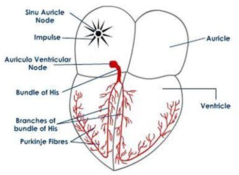
Structures Related to Origin and Conduction of Heartbeat:
1. Sinu-Auricular Node (SA Node) – "Pacemaker":
-
It is a specially modified neuro-muscular tissue.
-
Located in the dorsal wall of the right auricle, just above the opening of the superior vena cava.
-
It initiates and regulates the heartbeat.
2. Auriculo-Ventricular Node (AV Node) – "Pace Setter":
-
Also a modified neuro-muscular tissue, but it cannot initiate impulses by itself.
-
It produces nerve impulses only after stimulation by the SA Node.
-
Located in the dorsal wall of the right auricle, near the auriculo-ventricular sulcus.
3. Bundle of His:
-
A bundle of specialized nerve fibers that originates from the AV Node.
-
It runs through the interventricular septum down to the apex (posterior part) of the heart.
-
At the posterior part, it bifurcates into right and left branches.
-
These branches further divide into fine, thread-like nerve fibers called Purkinje fibers, forming a net-like structure in the walls of both ventricles.
Origin and Conduction of Heartbeat
-
Heartbeat is originated in the form of a nerve impulse, which is followed by the contraction of heart muscles.
-
The heartbeat originates from the SA Node and is transmitted to the walls of both auricles, causing auricular systole.
-
The nerve impulse produced by the SA Node stimulates the AV Node, which then generates a nerve impulse for the systole of ventricles.
-
The nerve impulse generated by the AV Node is transmitted to the walls of both ventricles through the Bundle of His and Purkinje fibers, resulting in ventricular systole.
Cardiac Cycle
-
The phenomenon of heartbeat occurs at regular intervals and is repeated continuously.
-
This repeating process is called the cardiac cycle.

-
It includes the following phases:
1. Auricular Systole
-
Both auricles receive the nerve impulse from the SA Node and undergo contraction (systole) to pump blood into their respective ventricles.
-
This phase is called auricular systole and takes about 0.1 seconds.
Delay Time:
-
The time taken by the AV Node to generate a nerve impulse after stimulation by the SA Node.
-
It is about 0.1 seconds.
2. Ventricular Systole
-
Both ventricles contract after receiving nerve impulses from the AV Node via the Bundle of His and Purkinje fibers.
-
This phase is called ventricular systole and takes about 0.3 seconds.
-
During this phase:
-
The left ventricle pumps oxygenated blood to the aorta (systemic circulation).
-
The right ventricle pumps deoxygenated blood to the pulmonary artery (pulmonary circulation).
-
3. Joint Diastole
-
In this phase, both auricles and ventricles are relaxed (in diastole).
-
Called joint diastole or complete cardiac diastole.
-
This phase lasts about 0.4 seconds.
Duration of Heartbeat
-
A complete heartbeat takes approximately 0.8 seconds.
-
This means 70–72 heartbeats occur per minute.
Pacemaker
-
A pacemaker is a small device placed in the chest or abdomen to control abnormal heart rhythms.
-
It uses electrical pulses to prompt the heart to beat at a normal rate.
-
The sinu-auricular (SA) node is called the natural pacemaker of the heart.
Blood Pressure
-
Blood pressure is the force exerted by blood against the walls of arteries, veins, and capillaries.
-
It is of two types:
-
Systolic Blood Pressure: Maximum pressure during contraction (systole).
-
Diastolic Blood Pressure: Minimum pressure during relaxation (diastole).
-
-
Normal adult BP = 120/80 mm Hg
-
Range:
-
Systolic: 100 (lower limit) – 140 (upper limit)
-
Diastolic: 60 – 90
-
Problems Related to Blood Pressure
1. Hypertension
-
A condition where a person has consistently high blood pressure, e.g., 150/90 mm Hg.
-
Causes include:
-
Tension, fear, anxiety, obesity, stress, sorrow, emotional strain
-
Smoking, cholesterol-rich diet, nephritis, narrowing of arteries, and loss of arterial elasticity
-
2. Hypotension
-
A condition where a person has persistently low blood pressure, e.g., 100/50 mm Hg
-
Causes include:
-
Loss of blood
-
Heart's failure to pump efficiently
-
Heart Sounds
-
The sounds heard during valve closure in heartbeats are called heart sounds:
-
First sound (Lubb):
-
Produced by closing of AV valves (bicuspid and tricuspid)
-
Occurs at beginning of ventricular systole
-
-
Second sound (Dup):
-
Produced by closing of semilunar valves (aortic and pulmonary)
-
Occurs at the end of ventricular systole
-
-
Third heart sound:
-
Very low frequency, not audible via stethoscope
-
Occurs at the start of ventricular diastole
-
-
Fourth heart sound:
-
Occurs during the rapid filling of blood into ventricles
-
Also low frequency and often inaudible
-
-
Pulse
-
The wave of distension felt in the artery wall due to contraction of the left ventricle.
-
Can be felt by placing a finger on the wrist artery.
Heart Rate
-
The number of pulses per minute is called the heart rate.
-
Normal heart rate is about 72 beats per minute.
Electrocardiogram (ECG)
-
An ECG is the graphical representation of the electrical variations produced during the beating of the heart.
-
It records the electrical activity of the heart.
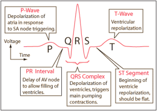
ECG Waves:
-
P wave – Depolarization of atria
-
QRS complex – Depolarization of ventricles
-
T wave – Repolarization of ventricles
✅ By counting the number of QRS complexes, the heart rate of an individual can be determined.
Clinical Interpretations:
-
Enlargement of P wave – Enlargement of atria
-
Enlargement of QR wave – Possible heart attack
-
S-T segment elevated – Indicates heart attack
-
Straight line (flat ECG) – Cardiac arrest or death of the patient
Stroke Volume (SV):
-
It is the volume of blood pumped out with every heartbeat.
-
Approximate volume: 70 ml
Cardiac Output (CO):
-
The volume of blood pumped by the heart in one minute.
-
Formula:
CO = Stroke Volume × Heart Rate
Blood Vessels:
Blood vessels are tubular structures that carry blood through the body.

Types:
-
Arteries:
-
Carry blood away from the heart under high pressure.
-
Carry oxygenated blood (except pulmonary artery).
-
Have thick walls and small lumen.
-
Composed of three layers:
-
Tunica interna: Thin elastic tissue
-
Tunica media: Smooth muscle
-
Tunica externa: Collagen and elastic fibers
-
-
-
Arterioles:
-
Small vessels that carry blood from arteries to capillaries.
-
-
Capillaries:
-
Very fine, thin-walled vessels that allow exchange of substances between blood and tissues.
-
-
Venules:
-
Small vessels that carry blood from capillaries to veins.
-
-
Veins:
-
Carry blood towards the heart under low pressure.
-
Carry deoxygenated blood (except pulmonary vein).
-
Have thin walls, large lumen, and valves to prevent backflow.
-
Arterial System:
-
Circulates oxygenated blood from the heart to the body via the aorta.
-
Aortic arch gives rise to:
-
Right Brachiocephalic Artery
-
Right common carotid
-
Right subclavian
-
-
Left Common Carotid Artery
-
Left Subclavian Artery
-
Descending Aorta branches:
-
Inferior phrenic → Diaphragm
-
Coeliac → Stomach and liver
-
Superior mesenteric → Small intestine
-
Renal → Kidneys
-
Suprarenal → Adrenal glands
-
Genital → Ovaries/Testes
-
Lumbar → Body wall
-
Inferior mesenteric → Large intestine
-
Common iliac → Legs
Venous System:
-
Carries deoxygenated blood back to the heart.
-
Superior Vena Cava:
-
Collects blood from:
-
Right & Left brachiocephalic veins
-
Jugular and subclavian veins
-
-
-
Inferior Vena Cava:
-
Collects blood from:
-
Common iliac veins
-
Lumbar, genital, renal, suprarenal, hepatic, inferior phrenic veins
-
-
Hepatic Portal System:
-
Blood from digestive organs flows to the liver before reaching the heart.
Constituent Veins:
-
Cystic (gall bladder)
-
Pancreatic (pancreas)
-
Gastric (stomach)
-
Duodenal (duodenum)
-
Superior mesenteric (small intestine)
-
Inferior mesenteric (large intestine)
-
Splenic (spleen)
Functions:
-
Stores excess glucose as glycogen
-
Converts ammonia to urea
-
Filters bacteria and toxins
-
Removes impurities
-
Regulates temperature
Blood Types (ABO System):
| Blood Group | Antigens in RBC | Antibodies in Plasma | Compatibility |
|---|---|---|---|
| A | A | Anti-B | Can receive A, O |
| B | B | Anti-A | Can receive B, O |
| AB | A and B | None | Universal recipient |
| O | None | Anti-A and Anti-B | Universal donor |
Rhesus (Rh) Factor:
-
Protein found on RBCs.
-
Rh positive = has the protein
-
Rh negative = lacks the protein
During Transfusion:
-
Rh-negative people receiving Rh-positive blood may develop antibodies, causing future transfusion reactions or complications.
During Pregnancy:
-
If Rh-negative mother carries an Rh-positive fetus, she may develop antibodies that harm future Rh-positive babies (hemolytic disease of the newborn).
Blue Baby Syndrome:
-
Caused by a hole in the ventricular septum → Mixing of oxygenated and deoxygenated blood.
-
Results in cyanosis (bluish skin/lips).
Heart Transplant:
-
First successful heart transplant: 3rd December 1967 by Dr. Christian Barnard in South Africa.
0 Comments
Post a Comment